Now readingIntroduction
0%
AP Biology · Framework 2026-27
Unit 1

Chemistry of Life

Biological structure begins with charge, bonding, and the controlled assembly of carbon-based molecules in water.

Topics
7

Introduction

Biological structure begins with charge, bonding, and the controlled assembly of carbon-based molecules in water.

This unit moves from water’s partial charges to the four major classes of biological molecules. The useful thread is structure: the location of charge changes molecular interactions, the order of subunits stores information, and the shape produced by those interactions constrains function.

Track atoms whenever molecules are joined or separated. Track interaction type whenever a structure changes. A covalent backbone, hydrogen bond, ionic attraction, and hydrophobic association do different jobs and respond differently to the environment.

01
Water polarity

aqueous environment and molecular interactions

02
Dehydration and hydrolysis

assembly and disassembly

03
Subunit order and side-chain chemistry

shape, information, and function

Current exam guidance: Aligned to the current 2026–27 AP Biology course framework, including scientific practices, data analysis, and model-based reasoning.
1.1

Structure of Water and Hydrogen Bonding

How does water’s molecular structure produce its biologically important properties?

Short answer

Water is polar, so neighboring molecules form temporary hydrogen bonds whose collective behavior produces cohesion, adhesion, thermal buffering, evaporative cooling, and surface tension.

Know firstElectronegativity · Polar covalent bonds · Partial-charge notation

Partial charges create a changing network

Oxygen attracts shared electrons more strongly than hydrogen. Because a water molecule is bent, its bond polarities do not cancel: oxygen carries a partial negative charge and the hydrogens carry partial positive charges. The partially positive hydrogen of one molecule can attract the partially negative oxygen of another. That intermolecular attraction is a hydrogen bond; it is not the covalent O–H bond inside a water molecule.

A single hydrogen bond is brief, but a sample contains an enormous, continually rearranging network. Energy added to liquid water changes molecular motion while also disrupting network contacts, so a given energy input produces a smaller temperature rise than it would in many comparison liquids. During evaporation, faster molecules are more likely to escape and carry energy away, lowering the average kinetic energy of the liquid left behind.

The same charge pattern explains cohesion between water molecules and adhesion between water and another polar surface. Cohesion supports surface tension because molecules at an interface experience an unequal inward attraction. Adhesion plus cohesion can help maintain a water column along a narrow polar surface. These effects are context-dependent; a nonpolar wall offers little adhesion even while water still coheres.

Teaching figure
Three-stage diagram connecting bent water molecules to temporary hydrogen bonds and collective network behavior

Partial charges connect water molecules

A water molecule’s bent shape leaves distinct partial charges. The attraction between unlike partial charges links neighboring molecules into a network that continually rearranges.

Use this visual to see
Follow the causal chain from molecular geometry to hydrogen bonding and then to bulk properties.
What it cannot show
The diagram shows a conceptual sequence, not molecular scale, bond lifetime, or a fixed number of neighbors.

Test Ninjas · Original educational diagram

Teaching figure
Comparison showing a liquid before evaporation, faster escaping molecules, and the cooler liquid remaining

Evaporation changes the molecules left behind

Molecules do not all move at the same speed. When faster molecules escape, they carry energy away and reduce the average kinetic energy of the liquid that remains.

Use this visual to see
Connect evaporative cooling to a change in the distribution of molecular speeds.
What it cannot show
The panels do not quantify an evaporation rate or show every collision at the liquid surface.

Test Ninjas · Original educational diagram

From charge to property

Polar charge distribution creates reversible attractions. Many attractions together resist rapid temperature change and produce cohesive behavior.

From charge to propertyA molecular ledger connects partial charges to hydrogen bonding, then connects the shifting network to thermal buffering and cohesion.
cooler
relativeMotion
1
averageHydrogenBondContacts
more
warmer
relativeMotion
2
averageHydrogenBondContacts
fewer and shorter-lived

Entities

  • bent water molecules
  • partial-positive hydrogen
  • partial-negative oxygen
  • thermal energy

Rules

  • Dashed links represent intermolecular hydrogen bonds, not covalent bonds.
  • Links continually break and reform.
  • Escaping molecules carry matter and thermal energy out of the liquid.
From charge to property states
StateidrelativeMotionaverageHydrogenBondContacts
1cooler1more
2warmer2fewer and shorter-lived

Use equal heating to compare temperature change

Which liquid in the synthetic comparison changes temperature less under the same energy input?

Use equal heating to compare temperature change dataset
LiquidStarting temperature (°C)Final temperature (°C)Change (°C)
water2127.26.2
comparison liquid2134.613.6
Provided accessible table
Use equal heating to compare temperature change accessible table
LiquidStart °CFinish °CChange °C
water2127.26.2
comparison liquid2134.613.6

Keep these three ideas

  • Bent geometry makes water polar.
  • Intermolecular hydrogen bonds explain several bulk properties.
  • Property claims require controlled comparisons, not a molecular cartoon alone.
1.2

Elements of Life

How do available elements constrain the molecules organisms can build?

Short answer

Cells rearrange conserved atoms into molecules: carbon supplies versatile backbones, while hydrogen, oxygen, nitrogen, phosphorus, and sulfur contribute characteristic groups and interactions.

Know firstAtoms and ions · Covalent bonding · Conservation of matter

Atoms are rearranged, not created

Carbon, hydrogen, and oxygen occur throughout biological molecules. Carbon can form several stable covalent bonds, so it supports chains, branches, and rings. Oxygen and nitrogen often create polar regions or participate in hydrogen bonding, while phosphorus commonly appears in nucleotide phosphate groups and phospholipid heads. Sulfur occurs in some amino-acid side chains and can help stabilize a folded protein.

The useful habit is atom accounting. An organism obtains elements from its environment in molecules or ions, breaks some bonds, and forms others. Photosynthesis can move carbon from carbon dioxide into carbohydrate, and cellular respiration can return carbon to carbon dioxide; neither pathway creates or destroys carbon atoms.

CHNOPS is a compact inventory, not a formula for every biological molecule. A carbohydrate need not contain nitrogen. A phospholipid contains phosphorus, but many other lipids do not. Many proteins contain sulfur, but a protein assembled without sulfur-containing amino acids is still a protein. Trace elements can be essential even though they are not major macromolecular building blocks.

Teaching figure
Three-panel comparison of carbon chain, branched, and ring frameworks

Carbon supports chains, branches, and rings

Carbon’s bonding capacity permits several stable backbone shapes. Those frameworks provide different places for functional groups and interactions.

Use this visual to see
Compare the backbone arrangements carbon can support without treating shape alone as proof of function.
What it cannot show
The symbols omit bond angles, electrons, hydrogens, and three-dimensional conformation.

Test Ninjas · Original educational diagram

Read composition without overgeneralizing

Molecule or componentElements shownWhat the composition supports
glucose, C₆H₁₂O₆C, H, Oa carbon-based carbohydrate example
generic sulfur-free amino acidC, H, O, Namino acids include nitrogen; sulfur is not universal
nucleotideC, H, O, N, Pphosphate and nitrogenous base are distinct components
phospholipidC, H, O, P and often other elementsan amphipathic lipid; not a template for every lipid

A closed carbon ledger

Enzyme-controlled reactions transfer carbon atoms through intermediates while conserving the number of atoms in the modeled system.

A closed carbon ledgerSix carbon atoms enter the model and six carbon atoms leave in a different molecular arrangement.
input
carbonAtoms
6
location
carbon dioxide pool
output
carbonAtoms
6
location
carbohydrate-equivalent pool

Entities

  • six carbon atoms in carbon dioxide
  • six carbon atoms in a carbohydrate product
  • reaction intermediates

Rules

  • Every incoming carbon receives an outgoing location.
  • Bond rearrangement may change molecular identity without changing atom count.
A closed carbon ledger states
StateidcarbonAtomslocation
1input6carbon dioxide pool
2output6carbohydrate-equivalent pool

Keep these three ideas

  • Carbon supports varied covalent frameworks.
  • Elemental composition constrains structure but does not determine function by itself.
  • Conservation requires every atom to be accounted for across a reaction.
1.3

Introduction to Macromolecules

How do cells join smaller molecules and later separate them?

Short answer

Dehydration forms a covalent connection while releasing the equivalent of water; hydrolysis adds water across a bond to separate the connected components.

Know firstCovalent bonds · Water · Conservation of atoms

Follow the bond and the water

In a simplified dehydration reaction, an H associated with one reactant and an OH associated with another leave as water while a new covalent bond connects the remaining groups. Hydrolysis reverses the atom placement: water contributes H and OH as the connecting bond is cleaved. The terms describe chemical accounting, not an organism becoming dehydrated or a molecule merely dissolving.

For one unbranched chain made from n separate monomers, joining the monomers requires n − 1 new connections and releases n − 1 water equivalents. Hydrolyzing every connection consumes the same number of water equivalents. This count changes when a problem specifies branching, ring closure, or several products, so first identify the topology rather than applying the expression blindly.

Cells use enzymes and energy coupling to control these reactions. A reaction label alone says nothing about rate or spontaneity. Lipids also mark an important boundary: many lipids are assembled from subcomponents, but they are not generally polymers made from one repeating monomer.

Teaching figure
Three-stage diagram of two monomers, dehydration forming a bond and water, and hydrolysis reversing the connection

Track the bond and the water molecule

Dehydration joins components while accounting for H and OH in water. Hydrolysis adds those atoms back across the bond that is cleaved.

Use this visual to see
Audit atoms while distinguishing assembly from disassembly.
What it cannot show
The diagram is a stoichiometric sketch and does not show an enzyme mechanism or reaction energetics.

Test Ninjas · Original educational diagram

Formula

Linear-chain bond count

b=n−1b=n-1

b is the number of new bonds, and therefore the number of water equivalents released, when n separate monomers form one open linear chain.

Use when
The model begins with separate monomers and ends with one unbranched, noncyclic chain.
Do not assume
A ring-closing bond, branch accounting, or multiple products changes the simple topology.

Check the count before applying it

Starting monomersNew linear-chain bondsWater released in dehydrationWater consumed in complete hydrolysis
2111
3222
6555

Keep these three ideas

  • Dehydration connects components and releases water equivalents.
  • Hydrolysis uses water to split the connection.
  • Count bonds from the stated topology and conserve every atom.
1.4

Carbohydrates

Why can carbohydrates serve both storage and structural roles?

Short answer

Monosaccharide identity, covalent linkage pattern, and branching change how a carbohydrate packs, how many accessible ends it has, and which enzymes can hydrolyze it.

Know firstTopic 1.3 dehydration and hydrolysis · Monomer and polymer language

Topology changes use

Monosaccharides can be joined by dehydration reactions into linear or branched polysaccharides. The resulting covalent linkage pattern is part of the structure, not a decorative detail: it affects chain shape, packing, and enzyme access. Hydrolysis can later release smaller sugars for other reactions.

Starch and glycogen are storage carbohydrates. Their glucose subunits contain chemical potential energy, and enzymes can expose or release subunits for metabolism. Branching can create multiple accessible chain ends. Cellulose, by contrast, aligns into strong fibers that contribute to plant cell-wall structure. The same broad monomer category can therefore support different functions when the linkages and larger arrangement differ.

A hexagon in a classroom diagram is only a symbolic subunit. The AP scope does not require memorizing detailed polymer structures. Avoid reducing every carbohydrate to immediate fuel: structural carbohydrates, short sugars, and storage polymers have different roles.

Teaching figure
Comparison of starch, glycogen, and cellulose organization and function

The same broad subunit can support different jobs

Storage and structural carbohydrates differ in linkage and larger organization. Branching changes end access, while aligned chains can support strong fibers.

Use this visual to see
Relate carbohydrate topology to storage access or structural support.
What it cannot show
The figure does not show detailed glycosidic-bond geometry and cannot identify a polymer from branching alone.

Test Ninjas · Original educational diagram

Same subunit count, different arrangement

Branching changes end availability, while repeated linear alignment can support fiber formation and structural strength.

Same subunit count, different arrangementThree arrangements use the same twelve symbols: one line, one four-ended branch, and parallel aligned rows.
linear
subunits
12
accessibleEnds
2
branched
subunits
12
accessibleEnds
4
aligned fibers
subunits
12
accessibleEnds
2
alignment
parallel

Entities

  • twelve symbolic monosaccharide subunits
  • linear chain
  • branched chain
  • aligned fiber bundle

Rules

  • Each state contains exactly twelve subunits.
  • Connections differ while matter count stays fixed.
  • Function is inferred from topology plus evidence, not topology alone.
Same subunit count, different arrangement states
StateidsubunitsaccessibleEndsalignment
1linear122—
2branched124—
3aligned-fibers122parallel

Connect named examples to a defensible function

CarbohydrateOrganization emphasizedMain AP-level useLimit
starchstorage polymer in plantsenergy reserveplant origin does not make every plant carbohydrate starch
glycogenhighly branched storage polymerenergy reserve in animals and fungibranching alone does not identify it
cellulosealigned structural polymerplant cell-wall supporthumans do not hydrolyze it to glucose with their own enzymes

Keep these three ideas

  • Monosaccharides can form linear or branched carbohydrates.
  • Linkage and arrangement influence access, packing, and function.
  • Starch, glycogen, and cellulose are examples, not interchangeable labels.
1.5

Lipids

How does lipid structure influence packing, assembly, and biological function?

Short answer

Hydrocarbon-rich regions avoid water, unsaturated tails usually pack less tightly, and amphipathic phospholipids orient into bilayers with heads toward water and tails inward.
Know firstPolarity and hydrophobic interactions · Molecular subcomponents

Hydrophobic regions organize in water

Lipids are grouped by physical behavior and function rather than as one repeating-monomer polymer family. Their hydrocarbon-rich regions interact poorly with water. In fats, fatty-acid tails with only carbon–carbon single bonds are saturated; one or more double bonds create unsaturation. In the standard biological model, bends associated with cis unsaturation disrupt close packing, often lowering the transition temperature relative to an equal-length saturated tail.

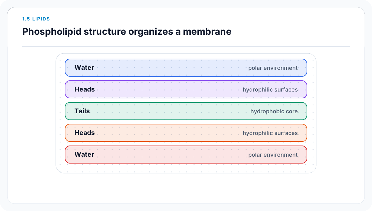
Phospholipids are amphipathic. Their polar heads interact with water while their nonpolar tails cluster away from it. In an aqueous environment this drives bilayer assembly: heads face the water on both sides and tails form the interior. The molecules are rearranged, not consumed. Cholesterol fits among phospholipids and helps stabilize animal membranes, but its effect depends on temperature and composition.

Other lipids have different jobs. Fats provide long-term energy storage and can insulate; steroids include signaling molecules. “Lipid” therefore does not mean “fat,” and “hydrophobic” does not mean every atom in every lipid avoids water. Chain length, double-bond geometry, temperature, and mixtures all limit a simple more-unsaturation-means-more-fluid rule.

Teaching figure
Layered membrane cross-section with water, polar heads, a nonpolar tail core, polar heads, and water

Water determines phospholipid orientation

Heads contact aqueous solutions on both sides while hydrocarbon tails gather inside. This arrangement creates the membrane’s chemically selective core.

Use this visual to see
Read a membrane from one aqueous face through its hydrophobic interior to the other face.
What it cannot show
The layers omit individual lipid motion, membrane proteins, cholesterol, and local curvature.

Test Ninjas · Original educational diagram

Bilayer orientation and tail packing

Hydrophobic association buries tails from water; bends disrupt orderly tail contact under matched conditions.

Bilayer orientation and tail packingA two-sided bilayer places round polar heads next to water and paired tails inside; a separate matched set shows lower illustrative transition temperatures with more unsaturation.
saturated
doubleBonds
0
relativePacking
tight
illustrativeTransitionTemperature
42
one unsaturation
doubleBonds
1
relativePacking
looser
illustrativeTransitionTemperature
28
two unsaturations
doubleBonds
2
relativePacking
loosest in this set
illustrativeTransitionTemperature
14

Entities

  • polar phospholipid heads
  • nonpolar tails
  • water on each membrane face
  • saturated and unsaturated comparison tails

Rules

  • Heads contact the aqueous regions.
  • Tails point toward the membrane interior.
  • The packing comparison holds tail length and temperature constant.
Bilayer orientation and tail packing states
StateiddoubleBondsrelativePackingillustrativeTransitionTemperature
1saturated0tight42
2one-unsaturation1looser28
3two-unsaturations2loosest in this set14

Keep lipid classes distinct

ClassStructural feature emphasizedBiological role
fatglycerol-associated fatty acidsenergy storage and insulation
phospholipidpolar head plus nonpolar tailsmembrane bilayer
steroidfour-ring carbon frameworksignaling and other functions
cholesterolamphipathic steroid lipidanimal-membrane stability

Keep these three ideas

  • Hydrophobic interactions help organize lipids in water.
  • Unsaturation changes packing under controlled conditions.
  • Amphipathic phospholipids form the membrane’s two-sided bilayer.
1.6

Nucleic Acids

How can a nucleic-acid strand store information and constrain the sequence of another strand?

Short answer

A directional sugar–phosphate backbone preserves base order, and specific complementary pairing lets one antiparallel strand serve as a template for another.

Know firstNucleotides · Hydrogen bonding · Covalent backbones

Sequence and direction carry the information

Each nucleotide contains a five-carbon sugar, phosphate group, and nitrogenous base. Covalent bonds join the sugar–phosphate backbone, leaving the base sequence available for information. The two ends are chemically different and are labeled 5′ and 3′. During synthesis, new nucleotides are added to the growing strand’s 3′ end, so sequence must always be read with direction attached.

DNA usually contains deoxyribose and bases A, T, G, and C. Its two strands run antiparallel: when one is written 5′ to 3′, its partner runs 3′ to 5′. A pairs with T and C pairs with G through hydrogen bonding. The covalent backbone preserves each strand; hydrogen bonds connect paired bases across strands. Complementarity allows one sequence to constrain a partner without making the strands identical.

RNA uses ribose and U in place of T and is typically single stranded. “Typically” matters: RNA can fold and form paired regions. A ladder diagram is also a model, not the literal geometry of a double helix. Pairing establishes sequence correspondence, but biological meaning depends on later replication, expression, and regulation.

Teaching figure
Antiparallel DNA strands showing template bases, complementary pairing, and opposite five-prime to three-prime directions

Direction is part of a DNA sequence

The sequence 5′-ACGTTA-3′ pairs with 3′-TGCAAT-5′. Writing both directions prevents a correct set of bases from being reported in the wrong order.

Use this visual to see
Build a complementary strand while keeping its orientation explicit.
What it cannot show
The straight strands do not represent helix geometry, grooves, scale, or replication enzymes.

Test Ninjas · Original educational diagram

Teaching figure
Three-stage diagram labeling phosphate, five-carbon sugar, and nitrogenous base

A nucleotide carries both backbone and sequence information

Phosphate and sugar participate in the directional backbone. The base is the variable component whose order stores information.

Use this visual to see
Separate the three nucleotide components and the role each plays.
What it cannot show
The simplified parts omit atom-level structure and the covalent bonds joining neighboring nucleotides.

Test Ninjas · Original educational diagram

Build the antiparallel partner

Complementary base selection constrains the partner sequence, and antiparallel orientation determines how that partner is written.

Build the antiparallel partnerThe template 5′-ACGTTA-3′ pairs with 3′-TGCAAT-5′; reversing that partner for standard reading gives 5′-TAACGT-3′.
template
orientation
5′ to 3′
sequence
ACGTTA
paired
orientation
3′ to 5′
sequence
TGCAAT
partner readable
orientation
5′ to 3′
sequence
TAACGT

Entities

  • template 5′-A C G T T A-3′
  • complementary bases
  • sugar–phosphate backbones

Rules

  • A pairs with T and C pairs with G.
  • Paired strands have opposite orientation.
  • Backbone links are covalent; base-pair links are hydrogen bonds.
Build the antiparallel partner states
Stateidorientationsequence
1template5′ to 3′ACGTTA
2paired3′ to 5′TGCAAT
3partner-readable5′ to 3′TAACGT

DNA and RNA: useful distinctions

FeatureDNARNA
sugardeoxyriboseribose
base used in A pairingthymineuracil
usual strand organizationdouble strandedsingle stranded with possible folded paired regions
shared featuredirectional nucleotide polymerdirectional nucleotide polymer

Keep these three ideas

  • Nucleotide order stores information in a directional polymer.
  • DNA strands pair complementarily and antiparallel.
  • Backbone and base-pair bonds perform different structural jobs.
1.7

Proteins

How does amino-acid sequence constrain protein shape and function?

Short answer

Amino-acid order places chemically different side chains in a specific pattern; backbone hydrogen bonding and side-chain interactions then favor a context-dependent three-dimensional shape that enables or limits function.
Know firstAmino-acid structure · Noncovalent interactions · Dehydration synthesis

Sequence establishes the interaction possibilities

A protein begins as a polypeptide: amino acids joined by peptide bonds in a specific order, with an amino end and a carboxyl end. Every amino acid contributes the same backbone pattern but a different side chain. The sequence therefore distributes nonpolar, polar, charged, and chemically reactive groups along the chain.

Local backbone hydrogen bonds support recurring secondary structures. Interactions among side chains and with the surrounding environment drive further folding: hydrophobic groups often cluster away from water, opposite charges can attract, similar charges repel, and some side chains can form covalent disulfide links. Multiple polypeptides may then associate. These levels describe organization; a protein does not visit them as four independent assembly stations.

Shape influences binding, catalysis, transport, signaling, movement, and structure because only some surfaces have compatible geometry and chemistry. A sequence change can alter function strongly, weakly, or not detectably, depending on its position and context. Temperature, pH, and chemical conditions can disrupt interactions and reduce function without necessarily breaking peptide bonds.

Scientific source
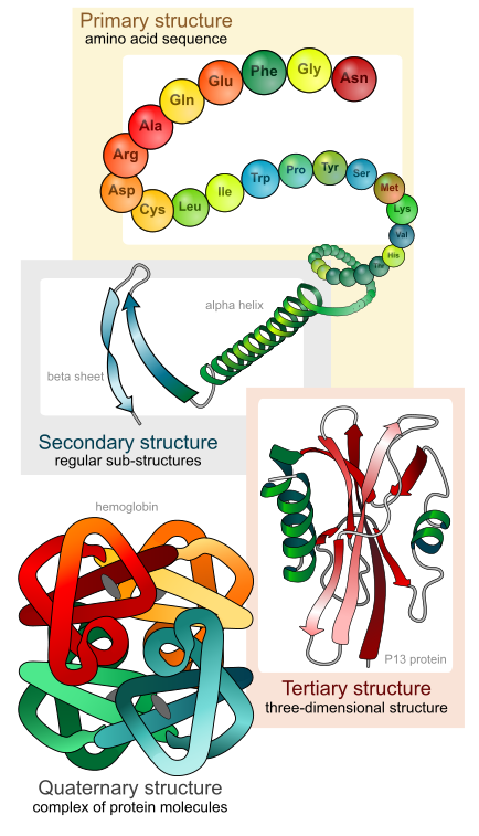
Public-domain diagram of primary, secondary, tertiary, and quaternary protein structure

Protein structure is organized at several scales

The levels describe different features of the same molecular system: sequence, local backbone patterns, one chain’s fold, and association among chains.

Use this visual to see
Locate what changes—and what remains the same—at each level of protein organization.
What it cannot show
The illustration presents representative shapes. Real proteins are dynamic, and not every protein has multiple subunits.

Mariana Ruiz Villarreal (LadyofHats) · Public domain · Wikimedia Commons item record

A side-chain perturbation changes one interaction network

Changing side-chain chemistry can remove a favorable contact, redistribute folding states, and alter the population able to bind a partner.

A side-chain perturbation changes one interaction networkThree synthetic states compare interaction count and activity. Removing one favorable charged contact lowers the modeled active fraction, while a same-class change has little effect.
reference
sequenceClass
N-H-P-N-H-P-C-N
favorableContacts
4
relativeActivity
1
charged to nonpolar change
sequenceClass
N-H-P-N-H-P-N-N
favorableContacts
3
relativeActivity
0.62
same class change
sequenceClass
N-H-P-N-H-P-C-N
favorableContacts
4
relativeActivity
0.96

Entities

  • eight-residue symbolic chain
  • hydrophobic side chains
  • positive and negative side chains
  • aqueous environment

Rules

  • Backbone order stays fixed unless a sequence change is specified.
  • Side-chain categories constrain favored contacts.
  • A folded-state drawing represents a population tendency, not a rigid molecule.
A side-chain perturbation changes one interaction network states
StateidsequenceClassfavorableContactsrelativeActivity
1referenceN-H-P-N-H-P-C-N41
2charged-to-nonpolar-changeN-H-P-N-H-P-N-N30.62
3same-class-changeN-H-P-N-H-P-C-N40.96

Organize protein structure without treating it as a staircase

LevelWhat is describedImportant stabilizing feature
primaryamino-acid sequencecovalent peptide bonds
secondarylocal backbone patternsbackbone hydrogen bonds
tertiaryone chain’s overall foldside-chain and backbone interactions
quaternaryassociation of multiple polypeptidesinteractions among subunits

Keep these three ideas

  • Sequence places side-chain chemistry in a specific order.
  • Many interactions produce a dynamic folded population.
  • Functional effect depends on where and how structure changes.

Chemistry of Life review

A change in charge, subunit order, or interaction can alter molecular shape; changed shape can alter binding, assembly, and biological function.

Recognize immediately

  • Separate covalent bonds within a molecule from attractions between molecules.
  • Conserve every atom through dehydration and hydrolysis.
  • Connect sequence or linkage pattern to three-dimensional behavior rather than naming a molecule alone.

Practice AP Biology in the real test engine.

Free practice set: timed, the same engine as our full-length tests, every answer explained.